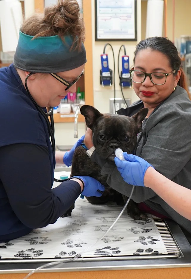
How often do we see an owner present a beautiful English Bulldog, Pug or similar brachycephalic puppy for a routine exam and vaccines and immediately notice that its nares are stenotic? Although the puppy is currently not showing any signs of respiratory difficulty, should we discuss upper airway disease with this owner who just purchased this beautiful puppy now, or should we wait until the pup starts to develop some of the typical clinical signs?
At the root of brachycephalic syndrome is the presence of increased upper airway resistance. This increased resistance is caused by narrowing of the nasal orifices (stenotic nares), an anatomical abnormality present at birth. Surprisingly, other areas of the upper airway do not initially have any abnormalities. English bulldog soft palates have been shown to have no appreciable anatomic changes or lesions at birth. Palates evaluated when clinical signs are present, however, show mucosal thickening, connective tissue edema, and mucous gland hyperplasia. It is suspected that these changes are secondary to constant exposure to increased stress placed on tissue due to increased negative airway pressure. Recent studies also have shown secondary abnormalities developing in turbinate associated tissue. This turbinate pathology causes even further narrowing of the airway passages resulting in a viscous cycle of increasing airway resistance and worsening airway obstruction. Essentially, because of the abnormal conformation, resistance to airflow is increased and any secondary soft tissue thickening exacerbates the clinical signs. For the patient, this increased airway resistance results in much more forceful inspiratory effort placing repetitive stress on laryngeal cartilage eventually leading to chondromalacia and progressive laryngeal collapse (a very different condition from laryngeal paralysis). Stage 2 and 3 laryngeal collapse in particular indicate end stage airway disease and tend to be poorly responsive to surgical treatment and carry a poor long term prognosis.
In addition to airway inflammation and obstruction, increased negative pressure exerted during inspiration is also suspected to lead to inflammatory changes in the intestinal tract. The pathophysiology behind these changes is poorly understood. One study suggested that virtually all brachycephalic dogs will have some degree of esophageal, gastric, or duodenal inflammation (or all of the above). When we delve into the history with many of these pet owners they will often report some form of gastrointestinal signs including dysphagia, gagging, and regurgitation. It is easy to dismiss gagging or “clearing of the throat” as directly related to the airway, but it seems that alimentary tract abnormalities likely contribute to these signs. One follow-up study showed a significant improvement in outcome if animals undergoing upper airway surgery were also started on medications to treat intestinal tract inflammation. In our hospital, we currently treat all routine brachycephalic airway cases with metoclopramide and famotidine and will avoid the administration of NSAIDS postoperatively. If intestinal signs are more pronounced, we will typically add a proton pump inhibitor and, in some instances, sucralfate to their perioperative and postoperative treatment regimen.
For the reasons outlined above, we strongly encourage early surgical intervention in dogs with brachycephalic syndrome in an attempt to minimize secondary changes in the airway simply by decreasing the degree and duration of high resistance to airflow as soon as possible. There are two approaches considered by many in these preclinical patients. A less aggressive surgical approach that should be considered is to perform correction of the patient’s stenotic nares at the time of spay or neuter at 4-6 months of age. This is a fairly minor procedure with a low risk for complications and high potential benefits if performed correctly. In many of these patients we will not yet see significant clinical changes in other parts of the airway. The disadvantage to this procedure is that the patient may require revision of their nares surgery as they mature. A second strategy is to delay sterilization in brachycephalic dogs until they are closer to skeletal maturity (10-12 months of age). At that time we will often see clinical evidence of early secondary changes such as elongation of the soft palate and eversion of laryngeal saccules and these issues will also need to be addressed when we correct their stenotic nares. In both situations early surgical intervention will hopefully prevent these patients from progressing to a point where they develop the much more serious complication of laryngeal collapse.
Long term outcome is usually good, especially if surgery is performed early in life. Owners need to be warned that improvement is usually instant (or occurs shortly after surgery) but that complete normalization of the airway is unlikely to be achievable and that some recurrence later in life (sometimes requiring revision of surgery) is possible. If laryngeal collapse is present, prognosis is less favorable and treatment options are few.
In our experience, clients with brachycephalic breeds tend to seek a surgical opinion only after clinical signs have advanced past the “normal” airway sounds and have led to exercise intolerance or an acute respiratory crisis. The acceptance of increased respiratory noise, stertor, stridor, and obesity as a state of normalcy in brachycephalic breeds by owners can be hugely detrimental to the long term outcome and quality of life of these animals. We truly believe that early intervention in these at-risk patients can result in significant improvement in long term quality of life for these patients and invite you to contact us at any time to discuss brachycephalic patients that you think may be at risk for developing (or are already showing) clinical signs so that we can help you devise the best possible plan for your patient and client.